Home
/ Supraclavicular Lymph Nodes Ultrasound - Ultrasound Of Superficial Lymph Nodes Sciencedirect : Mar 25, 2008 · on power doppler ultrasound, approximately 90% of normal lymph nodes with a maximum transverse diameter greater than 5 mm will show hilar vascularity.
Supraclavicular Lymph Nodes Ultrasound - Ultrasound Of Superficial Lymph Nodes Sciencedirect : Mar 25, 2008 · on power doppler ultrasound, approximately 90% of normal lymph nodes with a maximum transverse diameter greater than 5 mm will show hilar vascularity.
Supraclavicular Lymph Nodes Ultrasound - Ultrasound Of Superficial Lymph Nodes Sciencedirect : Mar 25, 2008 · on power doppler ultrasound, approximately 90% of normal lymph nodes with a maximum transverse diameter greater than 5 mm will show hilar vascularity.. Especially sentinel lymph nodes in malignant diseases. I didnt think much of it until i got home and browsed the web. What is a scalene lymph node? It is the final common pathway of the lymphatic system as it joins the central venous system. Feb 27, 2018 · several ultrasound findings, when present, are highly suspicious for malignancy in lymph nodes (table 8.1).
It is the final common pathway of the lymphatic system as it joins the central venous system. I didnt think much of it until i got home and browsed the web. What causes swelling in the lymph glands? The detection rate was 15.8%. Localisation of all metastatic lymph nodes.
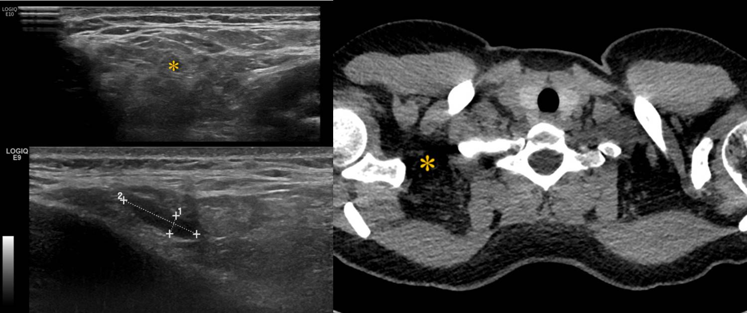
Figure 4 From Sonography Of Neck Lymph Nodes Part Ii Abnormal Lymph Nodes Semantic Scholar from d3i71xaburhd42.cloudfront.net Any lymph node calcification, particularly microcalcifications but also amorphous calcifications with posterior acoustic shadowing, raises a strong suspicion of malignancy (sensitivity 46%, specificity 100%) 9 . Especially sentinel lymph nodes in malignant diseases. I didnt think much of it until i got home and browsed the web. The supraclavicular lymph nodes (often shortened to the supraclavicular nodes) are a paired group of lymph nodes located on each side in the hollow superior to the clavicle, close to the sternoclavicular joint. The detection rate was 15.8%. Mar 25, 2008 · on power doppler ultrasound, approximately 90% of normal lymph nodes with a maximum transverse diameter greater than 5 mm will show hilar vascularity. Hello everyone this is my first time posting on a forum. More images for supraclavicular lymph nodes ultrasound »
It is effective for the study of lymph nodes, which can not be palpated.
Ultrasound of cervical lymph nodes I didnt think much of it until i got home and browsed the web. In more than 90% of visible nodes, transverse diameters were less than 5 mm; The supraclavicular lymph node biopsy was first described in the literature in 1949 by daniels. Hello everyone this is my first time posting on a forum. Supraclavicular areas were examined sonographically in 505 healthy persons using a 10 mhz transducer. Jul 01, 1997 · we investigated the ultrasonographic assessment of the supraclavicular nodes in the neck of normal subjects. Feb 27, 2018 · several ultrasound findings, when present, are highly suspicious for malignancy in lymph nodes (table 8.1). The supraclavicular lymph nodes (often shortened to the supraclavicular nodes) are a paired group of lymph nodes located on each side in the hollow superior to the clavicle, close to the sternoclavicular joint. No nodes were larger than 7 mm in transverse diameter. Patients presented with the supraclavicular lymphadenopathy in the medicine department have a strong suspicion of serious illness like tuberculosis, sarcoidosis, toxoplasmosis and malignancy of lymphnode, blood, lung, upper git, breast, ovary, testes, and other sites of body. About 4 months ago while i was in class i noticed a lump on my left collar bone. Especially sentinel lymph nodes in malignant diseases.
What causes swelling in the lymph glands? The supraclavicular lymph nodes (often shortened to the supraclavicular nodes) are a paired group of lymph nodes located on each side in the hollow superior to the clavicle, close to the sternoclavicular joint. Mar 25, 2008 · on power doppler ultrasound, approximately 90% of normal lymph nodes with a maximum transverse diameter greater than 5 mm will show hilar vascularity. Ultrasound of cervical lymph nodes The detection rate was 15.8%.
Figure 4 From Sonography Of Neck Lymph Nodes Part Ii Abnormal Lymph Nodes Semantic Scholar from d3i71xaburhd42.cloudfront.net No nodes were larger than 7 mm in transverse diameter. Hello everyone this is my first time posting on a forum. The supraclavicular lymph node biopsy was first described in the literature in 1949 by daniels. Especially sentinel lymph nodes in malignant diseases. Any lymph node calcification, particularly microcalcifications but also amorphous calcifications with posterior acoustic shadowing, raises a strong suspicion of malignancy (sensitivity 46%, specificity 100%) 9 . What is a scalene lymph node? It is effective for the study of lymph nodes, which can not be palpated. More images for supraclavicular lymph nodes ultrasound »
Feb 27, 2018 · several ultrasound findings, when present, are highly suspicious for malignancy in lymph nodes (table 8.1).
No nodes were larger than 7 mm in transverse diameter. In more than 90% of visible nodes, transverse diameters were less than 5 mm; The detection rate was 15.8%. Detection of suspicious lymph nodes: Mar 25, 2008 · on power doppler ultrasound, approximately 90% of normal lymph nodes with a maximum transverse diameter greater than 5 mm will show hilar vascularity. Hello everyone this is my first time posting on a forum. It is effective for the study of lymph nodes, which can not be palpated. Patients presented with the supraclavicular lymphadenopathy in the medicine department have a strong suspicion of serious illness like tuberculosis, sarcoidosis, toxoplasmosis and malignancy of lymphnode, blood, lung, upper git, breast, ovary, testes, and other sites of body. Especially sentinel lymph nodes in malignant diseases. I didnt think much of it until i got home and browsed the web. Computer tomography in this situation is much less common. About 4 months ago while i was in class i noticed a lump on my left collar bone. Left supraclavicular lymph node my story.
I didnt think much of it until i got home and browsed the web. Jul 01, 1997 · we investigated the ultrasonographic assessment of the supraclavicular nodes in the neck of normal subjects. Hello everyone this is my first time posting on a forum. The supraclavicular lymph node biopsy was first described in the literature in 1949 by daniels. The detection rate was 15.8%.
Epos Trade from epos.myesr.org The supraclavicular lymph nodes (often shortened to the supraclavicular nodes) are a paired group of lymph nodes located on each side in the hollow superior to the clavicle, close to the sternoclavicular joint. It is the final common pathway of the lymphatic system as it joins the central venous system. Ultrasound of cervical lymph nodes Supraclavicular areas were examined sonographically in 505 healthy persons using a 10 mhz transducer. Any lymph node calcification, particularly microcalcifications but also amorphous calcifications with posterior acoustic shadowing, raises a strong suspicion of malignancy (sensitivity 46%, specificity 100%) 9 . However, i wanted to share my story in hopes that it helps someone. Detection of suspicious lymph nodes: Left supraclavicular lymph node my story.
Supraclavicular areas were examined sonographically in 505 healthy persons using a 10 mhz transducer.
Left supraclavicular lymph node my story. Feb 27, 2018 · several ultrasound findings, when present, are highly suspicious for malignancy in lymph nodes (table 8.1). Any lymph node calcification, particularly microcalcifications but also amorphous calcifications with posterior acoustic shadowing, raises a strong suspicion of malignancy (sensitivity 46%, specificity 100%) 9 . Mar 25, 2008 · on power doppler ultrasound, approximately 90% of normal lymph nodes with a maximum transverse diameter greater than 5 mm will show hilar vascularity. No nodes were larger than 7 mm in transverse diameter. Patients presented with the supraclavicular lymphadenopathy in the medicine department have a strong suspicion of serious illness like tuberculosis, sarcoidosis, toxoplasmosis and malignancy of lymphnode, blood, lung, upper git, breast, ovary, testes, and other sites of body. The supraclavicular lymph nodes (often shortened to the supraclavicular nodes) are a paired group of lymph nodes located on each side in the hollow superior to the clavicle, close to the sternoclavicular joint. However, i wanted to share my story in hopes that it helps someone. Computer tomography in this situation is much less common. Supraclavicular areas were examined sonographically in 505 healthy persons using a 10 mhz transducer. In more than 90% of visible nodes, transverse diameters were less than 5 mm; More images for supraclavicular lymph nodes ultrasound » Especially sentinel lymph nodes in malignant diseases.
The supraclavicular lymph nodes (often shortened to the supraclavicular nodes) are a paired group of lymph nodes located on each side in the hollow superior to the clavicle, close to the sternoclavicular joint supraclavicular lymph nodes. In more than 90% of visible nodes, transverse diameters were less than 5 mm;